HAL® S3004
HAL® S3004 One Year Old Pediatric Simulator
Details:
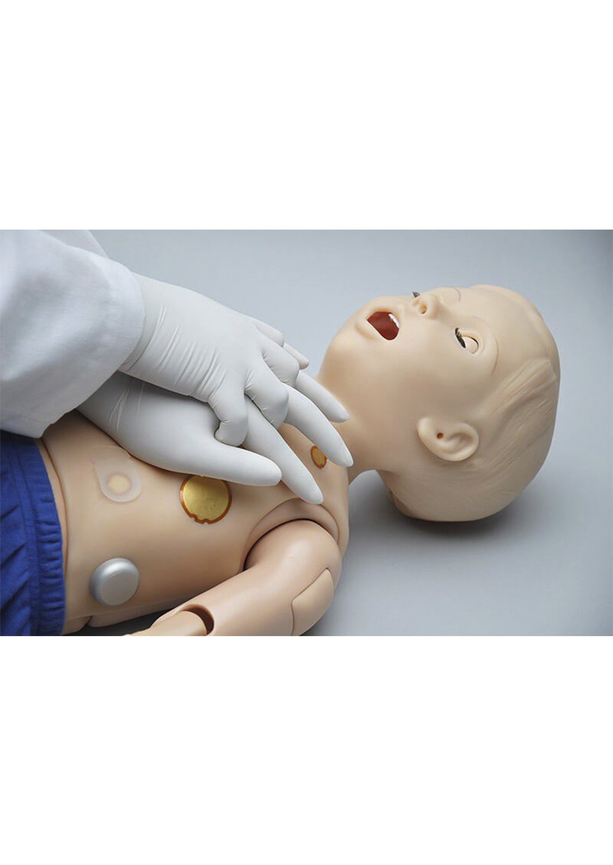
Pediatric HAL® allows you to take advanced simulation where you need to train. It may be at an accident scene, in an ER, an EMS vehicle, or even in a PICU. HAL® remains fully functional while being moved from place to place. This “Care in motion” allows you to evaluate both team training and how well patient “hand-offs” are conducted. What is done well? What needs to be improved?
Pediatric HAL® Allows You to Take Advanced Simulation Where You Need to Train.
|
Realistic
Realistic size and weight, tetherless connectivity, airway, chest rise, cyanosis, pre-recorded sounds and a variety of other features make for highly realistic scenarios.
|
Mobile
No external compressors, no linking boxes, no cords; just HAL® and a Tablet PC wirelessly connected for up to 300 feet.
|
Complete solution
From our standard one year warranty and pre installed scenarios, to multiple service, training, and warranty offerings, we cover all of your simulation needs.
|
Affordable
Gaumard® dedicates its talents to providing simulators at affordable prices. This principle remains as true today as it was over 60 years ago.
|
|
Intuitive software
Our intuitive and powerful user interface defines... Simulation Made Easy™
|
Debriefing
Evaluate interventions and insert notes on a real time performance log. Use an integrated camera system for comprehensive debriefing
|
Reliable
Standard one year warranty and over 60 years of experience building high quality patient simulators
|
Proven technology
Gaumard® pioneered wireless and tetherless simulators back in 2004. Pediatric HAL® is part of our growing family of these remarkable products.
|
 |
 |
 |
 |
|
Tetherless
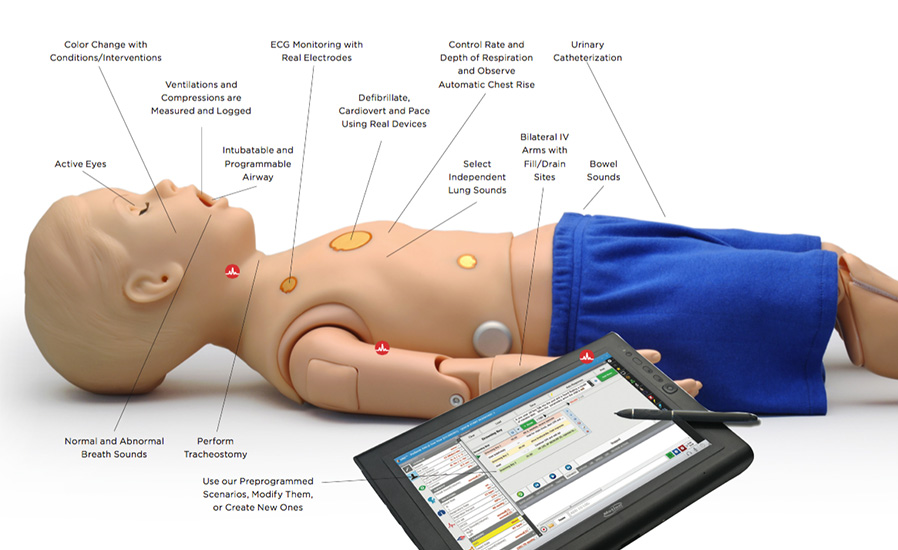
Control HAL® at distances up to 300 feet while he smoothly transitions between physiologic states in response to commands from a wireless tablet PC.
|
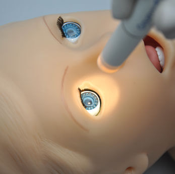
Active eyes
HAL® has blinking eyes with photo sensitive pupils. Dilation, reactivity, and blink rate can be controlled automatically or by the instructor.
|
Defibrillate, cardiovert and pace using real devices
HAL®’s electrically conductive skin regions allow the use of real equipment to obtain his ECG, perform temporary pacing, cardiovert, and defibrillate.
|
Airway & breathing
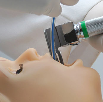
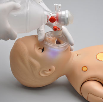
Improved airway allows better visualization of vocal cords and easy intubation. Lung compliance refined to deliver chest rise when ventilating at 20cm H2O.
|
 |
 |
||
|
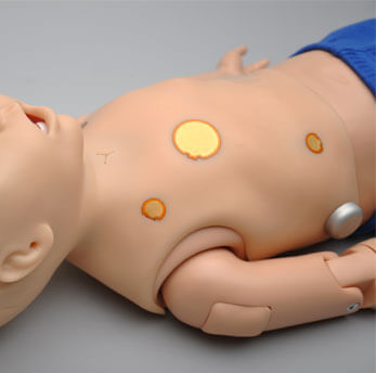
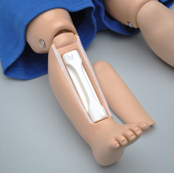
Intraosseous access
Intraosseous infusion and injection system with realistic tibia bones.
|
Cyanosis
Color and vital signs respond to hypoxic events and interventions.
|
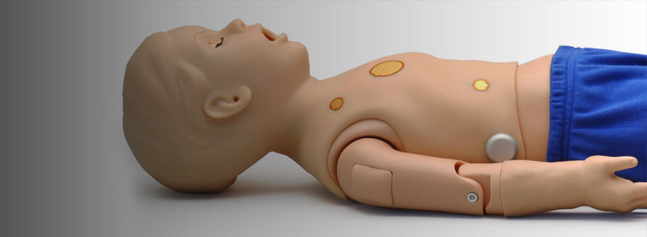 |
One year old
|
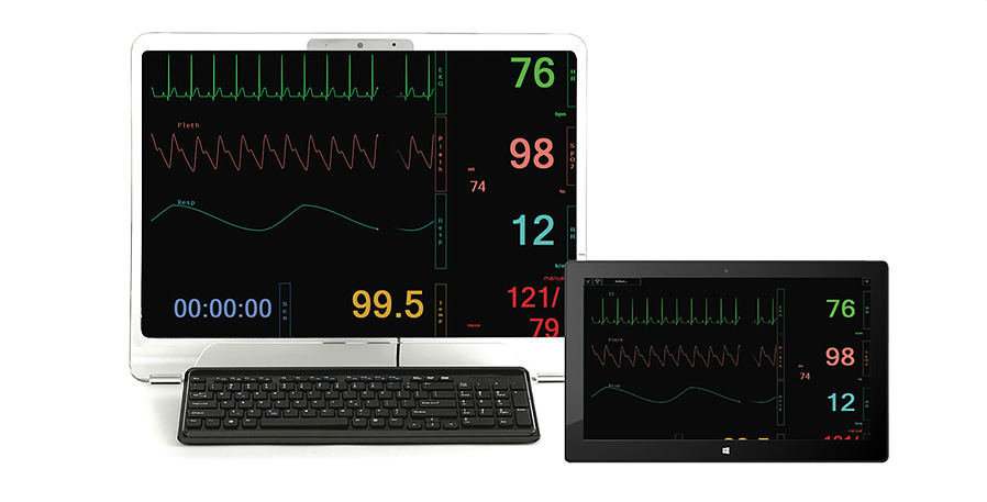
Monitoring
Touchscreen vital signs and perinatal monitors provide students with feedback provided in real clinical settings
Vital Signs for one-year-old HAL®
- Optional 20 inch “all-in-one” touchscreen virtual monitor AND a 12 inch touchscreen monitor
- Customize each trace independently; users can set alarms, and time scales.
- Display up to 12 numeric values including HR, ABP, CVP, PAWP, NIRP, CCO, SpO2, SvO2, RR, EtCO2, temperature, and time.
- Select up to 12 dynamic waveforms including ECG Lead I, II, III, aVR, aVL, aVF, V1, V2, V3, V4, V5, V6, AVP, CVP, PAWP, pulse, CCO, SvO2, respiration, capnography.
- Share images such as x-rays, CT scans, lab results, or even multimedia presentations as the scenario progresses.
Perinatal Monitor
- Dynamic Perinatal Monitors display uterine activity and fetal heart tones
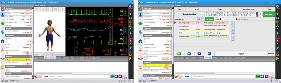
Pediatric HAL® S3004 - S3005 Features (07:00)
Standard features now include active eyes with programmable blink rate, pupil size and pupil reaction time as well as cyanosis and convulsions Resources Pediatric HAL® S3005 Manual Pediatric HAL® S3005...
Features
- Available in ethnic skin tones
- Tetherless and fully responsive even while being transported
- Powered from an internal rechargeable battery or wall outlet
- Simulator receives commands from a wireless tablet PC and operate at distances up to 300 feet
- Simulator can operate automatically using optional Automatic Mode or by the Instructor
- Training Guide with both basic and advanced interactive scenarios
- Use pre programmed scenarios, modify them or create your own quickly and easily
- Installation and training worldwide
- Simulation Made EasyTM
Airway
- Programmable airway
- Tongue edema
- Multiple upper airway sounds synchronized with breathing
- Nasal or oral intubation
- Right mainstem intubation
- Sensors detect depth of intubation
- Airway may be obstructed
- Block right lung, left lung, or both lungs
- Head tilt/ chin lift
- Suctioning techniques can be practiced
- Bag-Valve-Mask Ventilation
- Placement of conventional airway adjuncts
- Endotracheal intubation using conventional ETTs
- Retrograde intubation
- Sellick maneuver brings vocal cords into view
- Perform tracheostomy
- Tracheostomy care and suctioning
New Airway Features
- Realistic geometry and larger epiglottis. Better visualization of vocal cords as well as easy intubation
- Improved chest wall recoil during CPR
- Lung compliance refined to deliver chest rise when ventilating at 20cm H2O
Cardiac
- ECGs are generated in real time with physiologic variations never repeating textbook patterns
- Heart sounds may be auscultated and are synchronized with ECG
Breathing
- Control rate and depth of respiration and observe chest rise
- Automatic chest rise is synchronized with respiratory patterns
- Select independent left and right upper lung sounds
- Chest rise and lung sounds are synchronized with selectable breathing patterns
- Accommodates assisted ventilation including BVM and mechanical support
- Ventilations are measured and logged
- Gastric distension with excessive BVM ventilation
- Chest compressions generate palpable blood pressure wave form and ECG artifacts
- Detection and logging of ventilations and compressions
- Simulated spontaneous breathing
- Variable respiratory rates and inspiratory/expiratory ratios
- Bilateral chest rise and fall
- Unilateral chest rise simulates pneumothoraces
- Normal and abnormal breath sounds
Circulation
- Measure blood pressure by palpation or auscultation
- Use real modified BP cuff to measure blood pressure
- Korotkoff sounds audible between systolic and diastolic pressures
- Pulse sites synchronized with BP and heart rate
- Bilateral IV arms with fill/drain sites
- Realistic flashback
- SubQ and IM injection sites
- Intraosseous access at tibia
- Chest compressions are measured and logged
- ECG monitoring using real devices
- Defibrillate, cardiovert and pace using real devices
- Multiple heart sounds, rates and intensities
- ECG rhythms are generated in real time
- Heart sounds synchronized with ECG
- Dynamic rather than static 12 lead ECG display available with Automatic Mode
- Pacing may be practiced anteriorly to avoid having to roll the patient during delivery
- Bilateral carotid, radial, brachial and femoral pulses synchronized with ECG
- Pulses vary with blood pressure, are continuous and synchronized with the ECG even during a paced rhythm
Neural Responses
- Eyes are controlled automatically by physiologic model or directly by the Instructor
- Eyes open and close
- Select blink rate
- Select pupillary response to light
Speech
- Pre recorded sounds
- Optional wireless streaming audio
Articulation and Movement
- Seizure/convulsions
- Realistic rotation of the shoulder and hip joints
- Legs bend at the knees
- Supine or semi-recumbent positions
Other
- Central cyanosis
- Fill bladder and perform Foley catheterization
- Interchangeable genitalia
- Insert feeding tubes
- Remains fully functional even while in transit
- Bowel sounds
User Interface
- Sensors track student actions
- Changes in condition and care provided are time stamped and logged
- View the actions of up to 6 care providers using a responsive menu or write narrative
- Generate and share diagnostic lab results
- File sharing through Vital Signs Monitor
- Links with optional Pro+ recording and debriefing system integrating the event log with cameras and patient monitor
- Supplied with wireless tablet PC
- 12 pre programmed scenarios which can be modified by the instructor even during the scenario
- Create your own scenarios - add/edit